Breaking

Category: Health

  • How to Quit Vaping Without Gaining Weight

    How to Quit Vaping Without Gaining Weight

    You want to quit vaping — for your lungs, your wallet, your future — but you have heard the stories: everyone gains weight when they quit nicotine. And it is true that weight gain is the most common side effect of quitting, averaging 5-10 pounds in the first year. But it is not inevitable, and understanding why it happens gives you the tools to prevent it. Nicotine affects appetite, metabolism, and oral habits simultaneously; quitting removes all three effects at once. Here is how to quit vaping without the weight gain — or at least without meaningful gain.

    Why Quitting Nicotine Causes Weight Gain

    Three mechanisms drive post-quitting weight gain, and all are manageable once named. First, metabolic: nicotine artificially raises resting metabolic rate by roughly 7-10% — quitting removes that boost, meaning you burn 100-200 fewer calories daily doing nothing. The effect is real but modest — it accounts for maybe 3-5 pounds over a year, not 20. Second, appetite: nicotine suppresses hunger signals and dulls taste; quitting restores both, so food tastes better and hunger feels stronger — most quitters eat 200-300 extra calories daily without noticing. Third, the oral fixation: vaping occupied your mouth and hands hundreds of times daily; the behavioral void gets filled with snacking almost automatically — the hand-to-mouth habit transfers directly to food. Combined, the average quitter faces a 300-500 calorie daily surplus they never had before. The good news embedded in this: every mechanism is behavioral or modestly metabolic, which means every mechanism has a countermeasure. Weight gain after quitting is not mysterious or uncontrollable — it is predictable physics with known fixes. The Smokefree.gov quit resources address weight concerns in their quit planning.

    Use Nicotine Replacement Strategically (It Controls Both Problems)

    Here is the counterintuitive key: nicotine replacement therapy (NRT) — patches, gum, lozenges — does not just ease cravings; it directly prevents weight gain by maintaining the metabolic and appetite effects while you break the behavioral habit. Studies consistently show quitters using NRT gain significantly less weight than cold-turkey quitters, because the nicotine dose tapers gradually instead of vanishing overnight. The strategy: use combination NRT (patch for baseline plus gum or lozenges for cravings) for the full recommended 8-12 weeks, then taper the NRT itself slowly — this separates quitting vaping (behavioral) from quitting nicotine (pharmacological) into two manageable steps instead of one brutal one. Do not rush off NRT to “be truly quit” — extended NRT use is safe, and the weight data favors the slow taper. Prescription options (varenicline/Chantix, bupropion/Zyban) also reduce weight gain — bupropion in particular is associated with less post-quit weight gain and can be discussed with your doctor. The principle: manage the nicotine taper deliberately, and the weight problem shrinks dramatically. Our health guides cover more on navigating health behavior change.

    Pre-Plan Your Food Environment Before Quit Day

    The oral-fixation snacking is the most preventable weight-gain driver — but only if you plan before cravings hit. Stock the environment: crunchy vegetables (carrots, celery, bell peppers, cucumber) pre-cut and visible — the crunch satisfies the hand-to-mouth loop with ~35 calories per cup; sugar-free gum and mints for the oral fixation; sparkling water with lemon for the ritual of holding and sipping something; air-popped popcorn (100 calories for 3 cups) for evening cravings; frozen grapes or berries for sweet cravings. Remove the traps: the chips, cookies, and candy that turn mindless snacking into 500-calorie sessions — you cannot eat what is not there during a 9 p.m. craving. Plan structured meals and snacks: quitting on an erratic eating schedule amplifies hunger-driven overeating; three meals plus two planned snacks stabilizes the appetite nicotine used to suppress. Keep a simple food log for the first month — not to diet, but for awareness; most quitters are shocked to discover the extra 300 calories, and awareness alone curbs half of it. The goal during quitting is not weight loss — it is weight stability. Diet later; stabilize now.

    Move More (The Double-Duty Intervention)

    Exercise is the only intervention that simultaneously fights cravings and prevents weight gain. The craving mechanism: even 5-10 minutes of moderate activity measurably reduces nicotine cravings — it occupies the body, releases the dopamine and endorphins the brain misses, and breaks the cue-craving cycle. The weight mechanism: 30 minutes of brisk walking burns ~150 calories — roughly offsetting the metabolic drop from quitting — while preserving the muscle mass that keeps metabolism healthy. The practical protocol: schedule a walk for your known hardest craving times (after meals, the commute, the evening wind-down — map your vaping triggers and pre-assign walks to them); keep walking shoes visible as the craving response; add 2-3 weekly strength sessions if possible (muscle is metabolically expensive tissue — preserving it during quitting protects long-term weight). Start the exercise habit 1-2 weeks before quit day so it is established when cravings peak. Do not aim for heroic workouts — consistency of moderate movement beats intensity. And reframe: every craving you walk through is both a craving defeated and weight-gain prevented — the single most efficient health behavior available during quitting.

    Keep Perspective: The Math Favors Quitting

    Step back for the honest accounting, because weight fear keeps too many people vaping. The average post-quit gain is 5-10 pounds, mostly in the first 3 months, and it plateaus — it is not endless. The cardiovascular benefit of quitting nicotine dwarfs the risk of 10 pounds: quitting cuts heart disease risk roughly in half within a year, while 10 pounds raises it marginally — the net health effect is overwhelmingly positive. Most quitters who gain weight lose it in the second year once the behavioral dust settles and normal appetite regulation returns. And the quitters who gain the most are typically cold-turkey quitters with no plan — the NRT-plus-food-environment-plus-exercise approach described here keeps most people within 0-5 pounds. If weight does climb despite your efforts, address it after the quit is solid (3+ months) — trying to diet aggressively during acute quitting undermines both efforts; sequential goals beat simultaneous ones. The hierarchy is clear: quitting vaping with 8 pounds gained beats vaping indefinitely at a lower weight by every health metric that matters. Do not let the smaller fear protect the bigger harm. If you are planning your quit, tell us about it — and then set the date.

    Quitting vaping causes weight gain through three mechanisms: metabolic slowdown (~100-200 fewer daily calories burned), restored appetite, and oral-fixation snacking. Counter all three: taper with nicotine replacement (patch plus gum/lozenges), pre-stock crunchy low-calorie snacks and remove junk food, walk through cravings, and stabilize eating patterns. Expect 0-5 pounds with this plan versus 5-10 without — and remember quitting with slight gain beats vaping indefinitely.

    Conclusion

    Quitting vaping without gaining weight is entirely achievable with a plan: NRT to taper nicotine gradually, a pre-stocked food environment that channels oral fixation into vegetables and gum, daily walks that kill cravings and offset the metabolic dip, and structured meals that stabilize the returning appetite. Do not diet during quitting — stabilize, then address any remaining gain after three months. The weight fear is valid but manageable; the vaping harm is certain and ongoing. Set your quit date, stock the carrots, lace up the walking shoes, and take the win. Learn more about our practical health coverage. The American Lung Association’s quit resources include free coaching programs for quitters.

    Frequently Asked Questions

    How much weight will I gain when I quit vaping?

    Average is 5-10 pounds in the first year, mostly in the first 3 months. With nicotine replacement, planned snacking, and exercise, most people limit it to 0-5 pounds. Cold-turkey quitters with no plan gain the most.

    Does nicotine really speed up metabolism?

    Yes, modestly — about 7-10%, or 100-200 daily calories. It is real but small; the bigger weight-gain drivers are increased appetite and behavioral snacking. The metabolic effect alone accounts for only a few pounds yearly.

    Should I diet while quitting vaping?

    No — focus on weight stability, not loss, during quitting. Aggressive dieting plus nicotine withdrawal is a dual-deprivation setup that undermines both efforts. Stabilize through the quit (3+ months), then address any remaining weight with a proper plan.

    Does vaping cause weight loss?

    Nicotine suppresses appetite and slightly raises metabolism regardless of delivery method, so vapers may weigh slightly less than they would otherwise. But using vaping for weight control trades a few pounds for lung, cardiovascular, and addiction harm — an indefensible exchange.

    When does post-quit weight gain stop?

    It typically plateaus by 3-6 months as appetite regulation normalizes and the behavioral transition completes. Most quitters who gain weight lose some or all of it in the second year. Persistent gain beyond 6 months deserves its own diet and exercise plan.

  • What Foods Actually Help Lower Cholesterol, According to Cardiologists

    What Foods Actually Help Lower Cholesterol, According to Cardiologists

    Your cholesterol came back high, and you would rather change your diet than start a statin — or you are on a statin and want food to help, not just the pill. The internet is full of “cholesterol-busting superfoods” with exaggerated claims. Here is what cardiologists actually recommend: not miracle foods, but specific dietary components with real evidence for lowering LDL (the “bad” cholesterol), ranked by impact. No single food transforms your numbers; the pattern does. But some foods pull measurably more weight than others.

    Soluble Fiber: The Workhorse (Oats, Beans, Lentils)

    If cardiologists could prescribe one food category, it would be soluble fiber. The mechanism is elegant: soluble fiber forms a gel in the digestive tract that binds bile acids (made from cholesterol) and carries them out — forcing the liver to pull more LDL from the blood to make new bile. The dose-response is well established: every 5-10 grams of daily soluble fiber lowers LDL by roughly 5%. The champions: oats and oatmeal (a bowl provides ~4g), beans and lentils (6-8g per cup — the single densest source), barley, Brussels sprouts, avocados, and ground flaxseed. Practical target: work toward 10g+ of soluble fiber daily — oatmeal for breakfast plus a cup of beans or lentils somewhere in the day gets you there. The effect builds over weeks and requires consistency; fiber is not a supplement you take occasionally but a daily structural component of eating. Bonus effects: soluble fiber also blunts blood-sugar spikes and feeds beneficial gut bacteria. This is the least glamorous and most evidence-backed cholesterol intervention in nutrition — the foundation everything else builds on. The American Heart Association’s cholesterol guidance places fiber at the center of dietary management.

    Healthy Fats: Nuts, Olive Oil, and Fatty Fish

    The fat story has evolved past “all fat is bad” into a nuanced picture cardiologists are comfortable with. Nuts — almonds, walnuts, pistachios — lower LDL by 5-10% at about a handful (1.5 oz) daily; walnuts add omega-3 ALA, and the effect comes from unsaturated fats displacing saturated fats plus fiber and plant sterols. Olive oil (extra virgin) as your primary cooking and dressing fat: the Mediterranean pattern built on olive oil consistently shows the best cardiovascular outcomes in trials — it lowers LDL modestly while raising HDL function. Fatty fish (salmon, sardines, mackerel, trout) twice weekly: the omega-3s (EPA/DHA) primarily lower triglycerides rather than LDL, but triglycerides are part of the lipid panel that matters, and fish displaces red meat — a double win. What to reduce simultaneously: saturated fat (fatty red meat, butter, full-fat dairy, coconut oil, palm oil) — the single dietary factor most directly raising LDL. You do not need to eliminate these, but the exchange matters: every saturated-fat calorie replaced with unsaturated fat measurably improves the lipid profile. Think substitution, not just addition. Our health library covers heart-healthy eating patterns in depth.

    Plant Sterols, Soy, and the Supporting Cast

    Beyond the big three, several foods earn cardiologist endorsement for modest but real effects. Plant sterols and stanols (found in fortified spreads, some orange juices, and naturally in nuts and seeds): they block cholesterol absorption in the gut, lowering LDL ~10% at 2g daily — the most potent food-based LDL intervention after fiber, though usually via fortified products rather than whole foods. Soy protein (tofu, edamame, soy milk): modest LDL reduction (~3-5%), useful as a meat replacement that compounds the saturated-fat displacement benefit. Fruits rich in pectin (apples, citrus, berries): contribute soluble fiber plus polyphenols with vascular benefits. Garlic: small LDL effect in some trials, more consistent modest blood-pressure benefit — worth including for the overall cardiovascular package. Dark leafy greens: the nitrates support blood-vessel function, complementing the lipid improvements. Green tea: catechins show modest LDL-lowering in meta-analyses. None of these alone moves the needle dramatically — but stacked onto the fiber foundation and fat substitution, they contribute to a dietary pattern that can lower LDL 15-25% in motivated adherents, approaching low-dose statin territory for some people.

    The Portfolio Diet: Putting It Together

    Cardiologists familiar with the research often point to the “Portfolio Diet” — a specific eating pattern combining all the cholesterol-lowering components: 50g+ daily nuts, 20g+ soluble fiber (oats, beans, barley), 2g plant sterols, and 50g+ soy protein. In trials, strict adherents lowered LDL by ~30% — comparable to early-generation statins. You do not need to follow it rigidly to benefit; the principle is stacking: a day built as oatmeal with walnuts and berries (fiber + nuts), lentil soup with olive oil (fiber + healthy fat), salmon with Brussels sprouts (omega-3s + fiber), and an apple for a snack (pectin) hits every mechanism simultaneously. The Mediterranean and DASH patterns achieve similar results through slightly different compositions — the common thread is plants, fiber, unsaturated fats, and minimal saturated fat and ultra-processed food. What undermines all of it: the “I ate oatmeal so the bacon doesn’t count” fallacy — adding good foods without displacing bad ones dilutes the effect. The pattern is the intervention; individual superfoods are just marketing.

    Honest Expectations: Food, Statins, and Your Numbers

    Set realistic expectations to avoid the two failure modes: dismissing diet as useless, or rejecting medication that is needed. Excellent dietary change lowers LDL roughly 15-25% in committed adherents — meaningful, sometimes enough to avoid medication for borderline cases, and always beneficial alongside medication. But genetics matter enormously: familial hypercholesterolemia and strong genetic predisposition can keep LDL high regardless of diet — this is not dietary failure but biology, and statins exist precisely for this. Cardiologists’ actual position: diet first for borderline elevations, diet plus medication for high risk — never medication instead of diet, because statins do not fix the dietary patterns driving inflammation, blood pressure, and diabetes risk alongside cholesterol. Recheck lipids 3 months after dietary change (that is how long the full effect takes to manifest). And track the full picture: diet that lowers LDL also typically improves triglycerides, blood pressure, blood sugar, and weight — a package no pill replicates. Work with your doctor on targets based on your overall cardiovascular risk, not cholesterol alone. If nutrition questions persist, contact us.

    Cardiologists’ top cholesterol-lowering foods: soluble fiber (oats, beans, lentils — ~5% LDL drop per 5-10g daily), nuts and olive oil (unsaturated fats displacing saturated), fatty fish twice weekly, plant sterols, and soy. Stacked as a dietary pattern, these lower LDL 15-25% — but the pattern matters more than any single superfood, and genetics sometimes require medication regardless of diet.

    Conclusion

    Lowering cholesterol with food is real but unglamorous: daily soluble fiber, nuts and olive oil instead of saturated fats, fatty fish twice a week, and plenty of plants — sustained for months, not sprinkled occasionally. Expect a 15-25% LDL improvement with genuine commitment, recheck in three months, and work with your doctor on whether diet alone suffices or medication joins the plan. Food is the foundation; for some, it is also enough. Learn more about our evidence-based nutrition coverage. The NHLBI’s cholesterol management guide details the Portfolio diet approach used in trials.

    Frequently Asked Questions

    Can diet alone lower cholesterol enough to avoid statins?

    Sometimes — committed dietary change lowers LDL 15-25%, which suffices for borderline cases. But genetic factors can keep LDL high regardless of diet; your doctor assesses overall cardiovascular risk to decide whether medication is needed alongside diet.

    Are eggs bad for cholesterol?

    Less than once thought — dietary cholesterol affects blood cholesterol modestly in most people; saturated fat matters more. Most cardiologists consider an egg daily fine within an otherwise heart-healthy pattern, though individual responses vary.

    How long before diet changes show in cholesterol tests?

    About 3 months for the full effect — recheck lipids after 12 weeks of consistent dietary change. Testing sooner underestimates the benefit and discourages adherence.

    Do I need to give up red meat entirely?

    No — but reduce frequency and portion, choose lean cuts, and treat it as occasional rather than daily. The key mechanism is displacing saturated fat with unsaturated fats and fiber, not absolute prohibition.

    What about coconut oil — is it heart-healthy?

    No — despite marketing, coconut oil is ~90% saturated fat and raises LDL comparably to butter in trials. Cardiologists recommend olive oil and other unsaturated oils instead. The “healthy” reputation is not supported by lipid research.

  • How to Treat a Sprained Ankle at Home Without Going to Urgent Care

    How to Treat a Sprained Ankle at Home Without Going to Urgent Care

    You rolled your ankle on the stairs — it is swelling, it hurts to walk, and urgent care means a $200 bill and three hours in a waiting room. The good news: most ankle sprains are minor ligament stretches that heal perfectly well at home with proper care. The key is knowing which sprains are safe to manage yourself and executing the home treatment correctly — because “walk it off” is bad advice, but so is rushing to the ER for every twisted ankle. Here is the complete home treatment protocol and the exact signs that mean you need professional care.

    First 10 Minutes: Assess Before You Treat

    Before anything else, determine what you are dealing with. A sprain stretches or partially tears ligaments; a fracture breaks bone — and they can feel similar initially. Do the Ottawa Ankle Rules, the validated clinical screen: can you bear weight — take four steps — both immediately after the injury and now? Is there tenderness on the bony prominences (the tip of the fibula/lateral malleolus, the tip of the tibia/medial malleolus, the base of the 5th metatarsal on the outer foot, the navicular bone on the inner midfoot)? Can you feel a deformity, or is the foot pointing at an abnormal angle? If you cannot bear weight at all, if there is a visible deformity, if there is numbness in the foot, or if the pain is immediately severe and unrelenting (rather than building gradually), skip home treatment and get imaging — those are fracture/dislocation patterns. If you can hobble, the ankle looks aligned, and pain is moderate and lateral (the classic “rolled outward” inversion sprain), you are in home-treatment territory. When in doubt, urgent care with X-ray is the safe call — a missed fracture heals badly. The American Academy of Orthopaedic Surgeons’ sprain guide details this assessment.

    The RICE Protocol — Updated (It’s Now PEACE & LOVE)

    The classic RICE (Rest, Ice, Compression, Elevation) has been refined by sports medicine into PEACE & LOVE — and the updates matter. First 1-3 days (PEACE): Protect — avoid movements that cause pain, but do not fully immobilize; a light elastic bandage or brace for support, crutches if you cannot walk without limping. Elevate — ankle above heart level as much as possible for the first 48 hours; elevation is the most effective swelling reducer. Avoid anti-inflammatories initially — this is the big update: inflammation is the healing process, and suppressing it aggressively in the first 48-72 hours may impair tissue repair (use acetaminophen for pain instead of ibuprofen in the first couple of days). Compress — elastic bandage from toes upward, snug but not numbness-tight. Educate — understand that active recovery beats passive rest. Then (LOVE): Load gradually — start putting weight on it as pain allows, progressing from partial to full; pain is your guide, not your enemy. Optimism — psychological outlook measurably affects recovery. Vascularization — gentle pain-free movement and cardio (upper body, or stationary bike when tolerated) promotes blood flow and healing. Exercise — progressive rehab exercises as soon as pain allows (below).

    The Rehab Exercises That Prevent Chronic Ankles

    Here is what separates ankles that heal fully from ankles that stay weak and re-sprain forever: rehabilitation. Start when you can bear weight with mild discomfort (usually days 3-7). Phase 1 — range of motion: trace the alphabet with your toes (3x daily), gentle ankle circles, towel-assisted stretches pulling toes toward you. Phase 2 — strengthening: resistance band exercises in all four directions (push down, pull up, turn in, turn out), 3 sets of 15, daily; heel raises progressing from seated to standing to single-leg. Phase 3 — balance/proprioception (the critical phase most people skip): single-leg stands 30 seconds (progress to eyes closed), wobble board or pillow stands, single-leg mini-squats. The sprain damaged not just ligaments but the proprioceptive nerves telling your brain where the ankle is — without retraining, the ankle remains “blind” and re-injury rates soar. Continue balance work for 4-6 weeks minimum. Return to sport criteria: full pain-free range of motion, strength equal to the other side, ability to hop on the injured foot without pain or instability, and successful completion of sport-specific movements. Rushing back without rehab is how a $0 home sprain becomes a $5,000 chronic instability problem. More injury recovery guidance in our health section.

    What to Expect: The Healing Timeline

    Set realistic expectations to avoid both panic and impatience. Grade I (mild stretch, minimal swelling, can walk with mild pain): 1-2 weeks to normal activity, rehab for 2-4 weeks. Grade II (partial tear, significant swelling and bruising, painful walking): 3-6 weeks, with rehab extending 6-8 weeks. Grade III (complete tear, major swelling, instability, often cannot bear weight initially): 8-12+ weeks and usually needs professional evaluation — if you suspect this grade, get assessed rather than home-managing. Swelling and bruising often look alarming (the foot can turn impressive shades of purple as blood tracks downward with gravity) — dramatic bruising alone, without the red flags below, is normal and not dangerous. Pain should steadily improve day over day after the first 48 hours; a plateau or worsening after day 3-4 warrants reevaluation. Full ligament healing takes 6-12 weeks even when you feel fine at 3 — the rehab continuation matters after pain resolves, because the tissue is still remodeling. Respect the timeline: ligaments heal slowly and cannot be rushed, only supported.

    Red Flags: When Home Treatment Isn’t Enough

    Certain signs mean stop home treatment and get professional evaluation. Seek care promptly if: you cannot bear weight at all (four steps) — possible fracture; there is visible deformity or the joint looks misaligned; numbness, tingling, or color changes in the foot (circulation or nerve compromise); pain is severe and unrelenting from the moment of injury rather than building; swelling does not begin improving after 3-4 days of proper PEACE treatment; you heard or felt a distinct “pop” with immediate severe swelling (can indicate complete ligament rupture or fracture); the ankle feels unstable or gives way repeatedly after the initial healing; or this is a repeat sprain of the same ankle (chronic instability needs professional assessment and possibly bracing or physical therapy). Also get checked if you have diabetes, circulation problems, or take blood thinners — these change the risk calculus. Urgent care can X-ray, rule out fracture, provide proper bracing, and refer to physical therapy — a single visit that prevents months of dysfunction is money well spent. Do not let cost anxiety turn a treatable injury into a permanent weakness. If injury questions arise, contact us.

    Most ankle sprains heal well at home: assess first (can you take four steps? any deformity or numbness?), then PEACE & LOVE — protect, elevate, compress, avoid early anti-inflammatories, then gradual loading with rehab exercises emphasizing balance retraining for 4-6 weeks. Get imaging for inability to bear weight, deformity, numbness, or swelling that worsens after day 3-4.

    Conclusion

    A sprained ankle does not automatically mean urgent care — most are home-manageable with correct assessment, the updated PEACE & LOVE protocol, and diligent rehab (especially the balance work everyone skips). Respect the red flags, respect the timeline, and do the exercises after the pain fades — that is what separates a two-week inconvenience from a lifetime of weak ankles. Your ligaments will heal; give them the right conditions. Learn more about our practical health coverage. The American Physical Therapy Association’s injury resources guide ankle rehab progressions.

    Frequently Asked Questions

    Should I use ice or heat for a sprained ankle?

    Ice (or cold) for the first 48-72 hours to limit swelling — 15-20 minutes several times daily. Heat comes later, once acute swelling subsides, to relax surrounding muscles. Never heat a freshly swollen sprain; it increases bleeding into the tissues.

    Can I walk on a sprained ankle?

    Yes, as pain allows — modern guidance favors early gradual loading over prolonged rest. Use crutches or a brace if limping significantly, and progress weight-bearing as pain decreases. Complete non-weight-bearing beyond a day or two is outdated for most sprains.

    Why does my ankle keep re-spraining?

    Almost always skipped rehabilitation — the sprain damaged proprioceptive nerves, and without balance retraining the ankle stays “blind” to its position. A full 4-6 week balance program after healing drops re-injury rates dramatically. Chronic instability may need bracing or PT evaluation.

    How do I know if it’s sprained or broken?

    The practical screen: inability to bear weight for four steps, tenderness directly on bony prominences (not just soft tissue), visible deformity, or numbness suggest fracture. When uncertain, get an X-ray — a missed fracture heals poorly and the visit is worth it.

    Should I take ibuprofen for a sprain?

    Current sports-medicine guidance suggests avoiding NSAIDs in the first 48-72 hours since inflammation drives healing; use acetaminophen for pain instead. After the acute phase, NSAIDs are reasonable if needed. This is an evolution from old “ice and ibuprofen immediately” advice.

  • Why Do I Always Feel Tired Even After 8 Hours of Sleep?

    Why Do I Always Feel Tired Even After 8 Hours of Sleep?

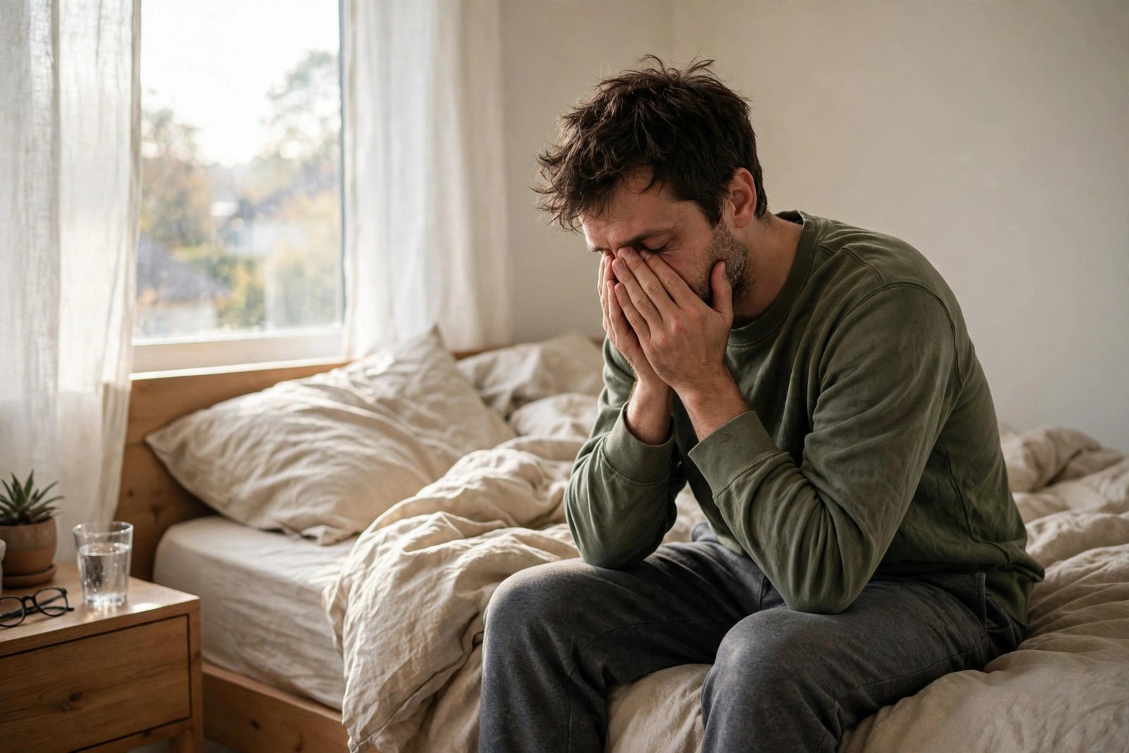
    You slept eight full hours — the supposedly magic number — and woke up feeling like you got four. The exhaustion is not just morning grogginess; it lingers through the day, coffee barely dents it, and by afternoon you are running on fumes. If this sounds familiar, you are experiencing one of the most common and most misdiagnosed complaints in medicine. The critical insight: sleep quantity and sleep quality are different things, and eight hours of poor-quality sleep restores less than six hours of good sleep. Here is why eight hours can leave you tired, and how to find your actual cause.

    The Core Distinction: Hours Asleep vs. Hours Restored

    Sleep is not a uniform state — it cycles through light sleep, deep slow-wave sleep, and REM sleep roughly every 90 minutes, and each stage does different restoration work. Deep sleep handles physical recovery, immune function, and growth hormone release; REM handles memory consolidation, emotional processing, and cognitive restoration. You can spend eight hours “asleep” while getting very little of either restorative stage — if something fragments your sleep architecture. The result has a specific feel: you wake unrefreshed, never quite reach full alertness, and experience daytime fatigue that sleep extension does not fix (sleeping 9-10 hours often makes it worse, not better). Fitness trackers’ sleep scores capture this imperfectly — total time looks fine while deep and REM percentages run low. The question is never just “how long did I sleep?” but “what kept my sleep shallow?” The causes below are ordered by how commonly they explain the eight-hours-but-exhausted pattern. The Sleep Foundation’s daytime sleepiness guide frames this quality-quantity distinction clinically.

    Cause #1: Sleep Apnea (The Most Missed)

    Obstructive sleep apnea is the leading medical explanation for unrefreshing sleep, and it is massively underdiagnosed — an estimated 80% of moderate-severe cases are undetected. The mechanism: your airway collapses repeatedly through the night, each collapse causing a micro-arousal (too brief to remember) that fragments sleep architecture. You can have dozens of these per hour — hundreds per night — while believing you “slept through.” The signature clues: loud snoring (ask your partner), witnessed breathing pauses, waking with headaches or dry mouth, needing to urinate multiple times nightly, and daytime sleepiness despite full nights in bed. Risk factors include excess weight, thick neck, male sex, and age — but thin people get it too. If this pattern fits, a home sleep test gives a definitive answer in one night, and CPAP treatment is often transformative — patients frequently describe it as the first real sleep in years. Do not accept “you just need more sleep” when eight hours is not working; get evaluated. Our health library covers sleep apnea diagnosis in detail.

    Cause #2: Fragmented Sleep You Don’t Remember

    Beyond apnea, numerous fragmenters destroy sleep quality invisibly. Alcohol is the most common self-inflicted one: it sedates you into unconsciousness but severely fragments the second half of the night — the classic pattern is falling asleep easily, then waking at 3 a.m. wired and unrefreshed. Even two drinks within 3-4 hours of bed measurably reduce restorative sleep. Medications: many antidepressants suppress REM sleep; beta-blockers can cause vivid dreams and fragmentation; antihistamines sedate but degrade architecture; diuretics taken too late cause bathroom awakenings. Environmental: bedroom noise you “sleep through” still triggers micro-arousals (traffic, snoring partner, HVAC cycling); light exposure (even dim light through eyelids suppresses melatonin); and temperature — a too-warm bedroom (above ~70°F) fragments sleep significantly. Restless legs syndrome and periodic limb movements cause hundreds of unconscious awakenings. Pain conditions (arthritis, back pain) fragment without full waking. The detective work: a two-week sleep diary noting alcohol, medications, bedroom conditions, and awakenings often reveals the fragmenter — or a partner’s observations do (“you toss all night,” “you stop breathing”).

    Cause #3: Circadian Misalignment

    You can sleep eight hours at the wrong biological time and wake exhausted. The circadian system expects sleep during the dark phase; sleeping against it — shift work, late-night screen habits pushing bedtime to 2 a.m., social jet lag (sleeping 1 a.m.-9 a.m. on weekends vs. 11 p.m.-7 a.m. weekdays) — produces sleep that is technically long enough but biologically mistimed. Misaligned sleep has less deep sleep, more fragmentation, and impaired next-day alertness even at equal duration. The modern epidemic version: the night-owl schedule forced into an early-bird world — sleeping midnight to 8 a.m. when your chronotype wants 2 a.m. to 10 a.m. produces chronic mild misalignment that feels exactly like “tired despite 8 hours.” Fixes: anchor your wake time (the single most powerful circadian signal — same time daily, weekends included); get bright light within 30 minutes of waking (outdoor light is 10x stronger than indoor); dim lights in the evening; and keep the sleep window consistent rather than “catching up” on weekends (which just reinduces jet lag weekly). If your schedule forces misalignment (shift work), strategic light exposure, melatonin timing, and sleep hygiene become medical necessities, not lifestyle tips.

    Cause #4: The Medical Causes Beyond Sleep

    Sometimes the tiredness is not primarily a sleep problem at all — the sleep is fine, but something else drains you. The common medical culprits: hypothyroidism (slows metabolism globally; fatigue is often the first symptom — a simple TSH test rules it out); iron deficiency, with or without anemia (impairs oxygen delivery; ferritin testing catches it — especially common in menstruating women); vitamin B12 deficiency (neurological fatigue, common in older adults and vegans); depression (fatigue and unrefreshing sleep are core symptoms — the tiredness of depression has a heavy, leaden quality distinct from sleepiness); chronic infections or post-viral syndromes; and sleep disorders beyond apnea (narcolepsy, idiopathic hypersomnia — rarer, but real). Medications deserve their own audit: statins, blood pressure drugs, antihistamines, and many others list fatigue as a side effect — review everything with your doctor or pharmacist. The workup for persistent unrefreshing sleep despite good habits is straightforward: thyroid panel, iron studies, B12, depression screening, medication review, and sleep apnea evaluation. Most causes are common, testable, and treatable — which makes persistent exhaustion without investigation the real problem.

    Your Diagnostic Action Plan

    Stop guessing and work the problem systematically. Week 1-2: run the sleep-hygiene fundamentals flawlessly — consistent wake time, morning bright light, no alcohol within 4 hours of bed, cool dark quiet bedroom, no caffeine after noon, screens off 30-60 minutes before bed. Track with a simple diary: bedtime, wake time, perceived quality, daytime fatigue (1-10), plus alcohol, caffeine, exercise. If exhaustion persists despite two genuinely good weeks: get the medical workup — primary-care visit with thyroid, iron/ferritin, B12, depression screening, and a sleep apnea risk assessment (bring your diary and, if possible, a partner’s observations about snoring and breathing pauses). Do not accept “you’re just stressed” without the basic labs — the treatable causes are too common. While investigating, resist the urge to simply sleep longer — extending time in bed with fragmented sleep usually worsens the unrefreshing pattern; protect sleep quality and consistency instead. And be patient with the process: the answer is usually in this list, and finding it changes everything. If persistent fatigue puzzles you, reach out — though your doctor runs the actual workup.

    Eight hours of tired means poor sleep quality, not insufficient quantity — something is fragmenting your sleep architecture or misaligning its timing. The usual suspects in order: sleep apnea (get tested — it is massively underdiagnosed), alcohol and other fragmenters, circadian misalignment from inconsistent schedules, and medical causes (thyroid, iron, B12, depression, medications). Run two weeks of strict sleep hygiene, then get the medical workup if exhaustion persists.

    Conclusion

    Feeling tired after eight hours is your body reporting that the sleep was not restorative — believe the report and investigate the cause rather than just adding hours. Fix the fundamentals, hunt the fragmenters, align your circadian rhythm, and get the straightforward medical workup if the basics do not resolve it. The answer is almost always findable and usually fixable; unrefreshing sleep is a symptom with a cause, not a life sentence. Learn more about our evidence-based health coverage. The American Thyroid Association’s patient resources explain thyroid-related fatigue and testing.

    Frequently Asked Questions

    Can you sleep too much and feel more tired?

    Yes — oversleeping (9-10+ hours regularly) is associated with grogginess and is often a marker of fragmented sleep: the body extends time in bed trying to compensate for poor quality. Fix quality and consistency rather than adding hours.

    How do I know if I have sleep apnea?

    Key signs: loud snoring, witnessed breathing pauses, morning headaches, dry mouth on waking, excessive daytime sleepiness despite adequate time in bed. A home sleep test gives a definitive answer in one night — ask your doctor.

    Does alcohol really ruin sleep quality?

    Yes — significantly. Alcohol sedates but fragments the second half of sleep, suppresses REM, and worsens apnea. Even moderate evening drinking measurably reduces restorative sleep; the effect is dose-dependent and worse closer to bedtime.

    What blood tests should I get for persistent fatigue?

    The standard panel: TSH (thyroid), ferritin and iron studies, vitamin B12, complete blood count, and depression screening. Your doctor may add others based on history. These catch the most common treatable causes.

    Is daytime fatigue the same as sleepiness?

    No — and the distinction guides diagnosis. Sleepiness (struggling to stay awake, dozing off) points toward sleep disorders like apnea. Fatigue (exhaustion without sleep tendency, heavy limbs) points more toward medical causes like thyroid, anemia, or depression. Note which one you feel.

  • How to Tell If Your Child Has an Ear Infection or Just a Cold

    How to Tell If Your Child Has an Ear Infection or Just a Cold

    Your child has a runny nose, a low fever, and has been tugging at their ear — or maybe just crying more than usual. Is it a cold that will pass, or an ear infection that needs a doctor? Parents face this question constantly, because colds and ear infections overlap heavily: most ear infections start as colds, and the early symptoms are nearly identical. Antibiotics do not help colds but are sometimes needed for ear infections, so telling them apart matters. Here is how to read the signs, what you can do at home, and when to call the pediatrician.

    Why Colds Turn Into Ear Infections

    Understanding the connection makes the distinction clearer. The middle ear — the small chamber behind the eardrum — connects to the back of the throat via the Eustachian tube, which equalizes pressure and drains fluid. In young children, this tube is shorter, narrower, and more horizontal than in adults, so it clogs easily. When a cold virus inflames the nasal passages and throat, the Eustachian tube swells shut, fluid gets trapped in the middle ear, and that warm, stagnant fluid becomes a perfect breeding ground for bacteria — turning a viral cold into a bacterial ear infection (acute otitis media). This is why ear infections so often follow colds by 3-5 days: the cold sets the trap, the bacteria spring it. Children under 5 are most susceptible (their anatomy is the least mature), and risk rises with daycare attendance, bottle-feeding while lying flat, pacifier use past 6 months, and exposure to tobacco smoke. Knowing this sequence — cold first, ear symptoms days later — is itself a diagnostic clue. The NIDCD’s ear infection resources explain this progression for parents.

    The Signs That Point to Ear Infection

    Several specific signs distinguish ear infection from uncomplicated cold. Ear pain is the hallmark — but young children cannot report it, so watch for: persistent ear tugging or pulling (occasional tugging is normal; relentless tugging with distress is not), crying that worsens when lying down (lying flat increases middle-ear pressure, intensifying the pain — babies who scream when put down but calm when held upright are telling you something), fever that spikes after the cold seemed to be improving (the classic pattern: cold for 3 days, then new fever of 102°F+ signals secondary bacterial infection), fluid draining from the ear (yellow, white, or bloody drainage means the eardrum may have perforated to release pressure — alarming to see, but it often relieves pain and usually heals on its own), trouble sleeping beyond normal cold fussiness, and loss of appetite or trouble swallowing (chewing and swallowing change middle-ear pressure). In older children who can talk, they will tell you directly: “my ear hurts” — believe them. The pattern to recognize: cold symptoms improving, then ear-specific symptoms and new fever appearing. That two-phase course is the ear infection signature.

    The Signs That Point to Just a Cold

    Equally important is recognizing when it is only a cold, because most colds never become ear infections and do not need medical visits. Points toward cold-only: runny nose and sneezing as the dominant symptoms (ear infections do not cause prominent nasal symptoms beyond the original cold); symptoms steadily improving day by day after the peak (around days 3-5); low-grade fever under 101°F that responds to acetaminophen or ibuprofen; the child is uncomfortable but consolable, sleeping reasonably, eating adequately; no new fever after initial improvement; no ear drainage; and ear tugging that is occasional and exploratory rather than distressed (babies discover their ears — not every touch is a symptom). Colds resolve in 7-10 days with supportive care: fluids, rest, saline nasal spray or suction for infants, humidified air, and honey for cough in children over 1 year. Antibiotics do nothing for colds and contribute to resistance — the reason pediatricians decline them is evidence-based, not dismissive. If the course is a straightforward cold getting better, home care is the right call. More on managing childhood illness in our health section.

    Home Care While You Watch and Wait

    For the uncertain middle — cold symptoms with possible early ear involvement — supportive care plus watchful waiting is the medically endorsed approach for most children over 6 months. Pain management: age-appropriate acetaminophen or ibuprofen (dosed by weight, not age — check the chart) relieves ear pain effectively regardless of cause; alternating them is unnecessary and complicates dosing. Warm compress on the affected ear (warm washcloth, 10-15 minutes) soothes pain for many children. Keep the child upright as much as comfortable — elevated head position reduces middle-ear pressure. Ensure fluids — dehydration worsens everything. Saline nasal spray plus gentle suction (infants) keeps the Eustachian tube end as clear as possible. What not to do: do not put anything in the ear canal (no oils, no drops, unless a doctor specifically directs — if the eardrum has perforated, liquids in the canal can cause harm); do not use leftover antibiotics from a previous illness (wrong drug, wrong dose, and it masks the clinical picture); do not demand antibiotics at the visit — guidelines support watchful waiting for 48-72 hours in many cases because most ear infections resolve without antibiotics. Document the course: when the cold started, when ear symptoms appeared, fever pattern, pain level — this history makes the pediatrician’s job far easier.

    When to Call the Pediatrician (and When It’s Urgent)

    Guidelines support watchful waiting, but certain situations need prompt evaluation. Call the pediatrician if: the child is under 6 months with any suspected ear infection (young infants are treated more aggressively); fever of 102.2°F (39°C) or higher with ear symptoms; severe ear pain not relieved by pain medication; ear drainage (especially bloody or foul-smelling); symptoms persisting beyond 2-3 days without improvement; the child seems unusually lethargic or irritable; or this is a recurrent pattern (3+ infections in 6 months or 4+ in a year — may warrant ENT referral for tubes). Seek urgent care if: the child is under 3 months with fever of 100.4°F+ (always urgent at that age); there is swelling, redness, or tenderness behind the ear (possible mastoiditis — a serious complication); the child cannot be awakened normally or is limp; or there are signs of dehydration (no tears, dry mouth, no wet diapers for 8+ hours). At the visit, the doctor will look at the eardrum with an otoscope — a red, bulging, immobile eardrum confirms infection; a normal eardrum rules it out definitively. This 30-second exam is why the visit matters: no home observation substitutes for seeing the eardrum. Trust the exam over the symptom guess. If parenting health questions persist, reach out to us.

    Colds cause runny noses and gradually improve over 7-10 days; ear infections typically follow colds by 3-5 days with new fever, worsening ear pain (worse lying flat), and sometimes drainage. Supportive care plus 48-72 hours of watchful waiting is appropriate for most children over 6 months — but infants under 6 months, high fever, drainage, severe pain, or recurrent infections warrant prompt pediatric evaluation. Only an otoscope exam of the eardrum confirms it.

    Conclusion

    The cold-versus-ear-infection question resolves with pattern recognition: steady improvement means cold; a second wave of fever and ear pain days into the cold means likely infection. Support the child through either, manage pain properly, watch and wait when appropriate, and let the pediatrician’s otoscope settle the uncertain cases. Most ear infections resolve well — with or without antibiotics — and the anatomy that makes toddlers susceptible matures with time. You are not expected to diagnose this at home; you are expected to recognize the patterns that merit a call. Learn more about our family health coverage. The American Academy of Pediatrics’ parent site has ear infection decision guides for parents.

    Frequently Asked Questions

    Does ear tugging always mean ear infection?

    No — babies tug their ears for many reasons including teething, exploration, and tiredness. Persistent, distressed tugging combined with other signs (fever, cold history, pain lying flat) is what suggests infection, not casual tugging alone.

    Will the eardrum heal if it ruptures from infection?

    Usually yes — most perforations from ear infections heal on their own within weeks once the infection resolves. The drainage often brings pain relief. Keep water out of the ear during healing and follow up as your pediatrician directs.

    Do all ear infections need antibiotics?

    No — guidelines support watchful waiting for 48-72 hours in many children over 6 months with mild symptoms, because most resolve spontaneously. Antibiotics are recommended for severe symptoms, very young infants, or cases that do not improve with observation.

    Can ear infections be prevented?

    Risk reducers: breastfeeding, avoiding bottle-feeding while flat, reducing pacifier use after 6 months, keeping up with vaccinations (flu and pneumococcal vaccines prevent many infections), avoiding tobacco smoke exposure, and good hand hygiene to reduce colds.

    When do recurrent ear infections need tubes?

    Generally after 3 infections in 6 months or 4 in 12 months, an ENT may recommend tympanostomy tubes — tiny tubes in the eardrums that ventilate the middle ear. It is a brief, common procedure with strong evidence for reducing recurrences.

  • What Is a Normal Resting Heart Rate for a Woman in Her 50s?

    What Is a Normal Resting Heart Rate for a Woman in Her 50s?

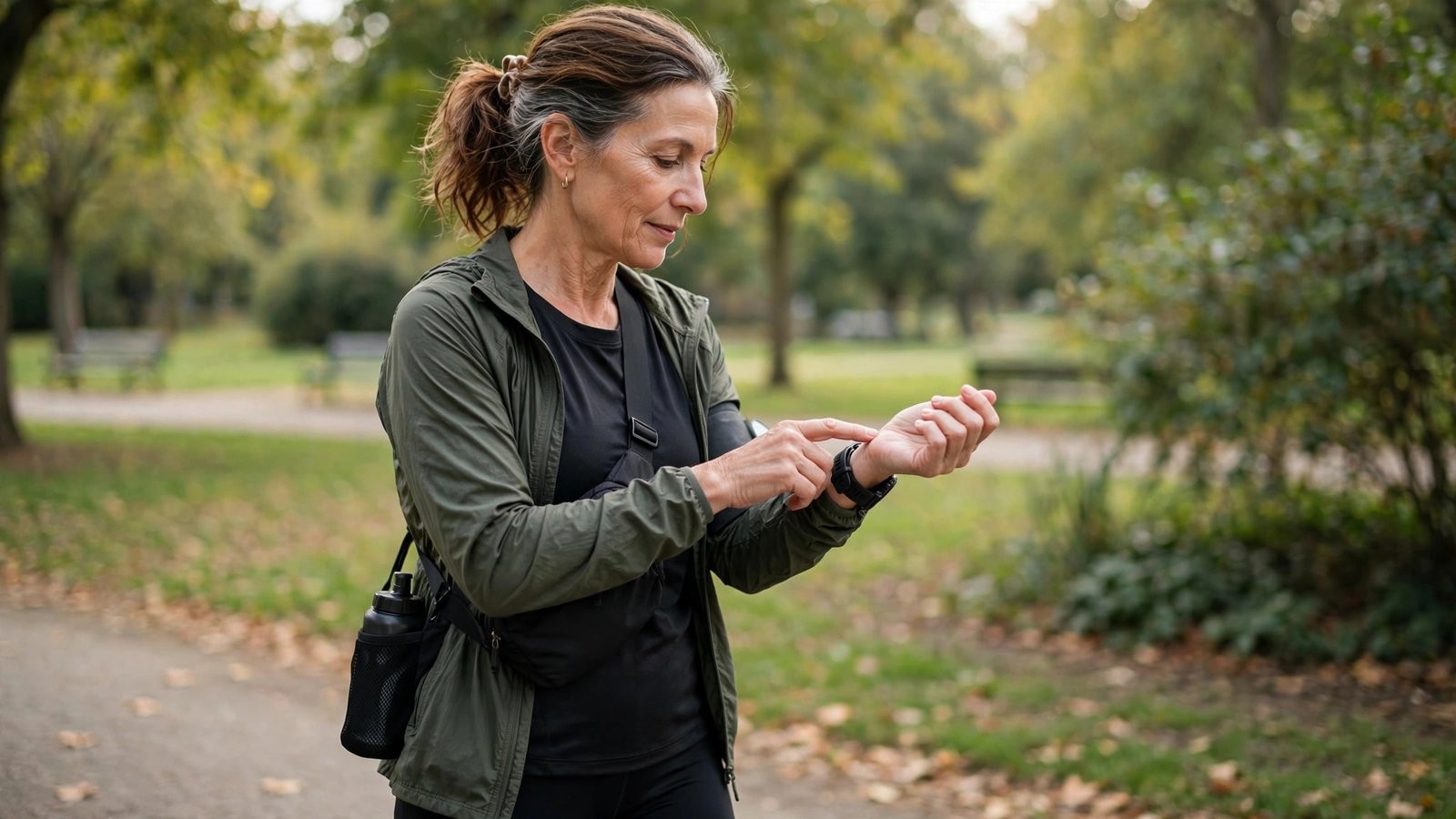
    You checked your pulse — maybe with a fitness tracker, maybe fingers on your wrist — and got a number: 72? 68? 81? Now you are wondering whether that is normal for a woman in her 50s, or a sign something is off. Resting heart rate is one of the simplest health metrics you can measure, and it carries real information about cardiovascular fitness — but “normal” has a wider range than most people think, and the number means little without context. Here is what is normal, what affects it, and when to pay attention.

    The Normal Range (Wider Than You Think)

    For adults, the clinically normal resting heart rate spans 60-100 beats per minute — and that range applies to women in their 50s as it does to everyone else. There is no separate “normal” for 50-something women; the range does not meaningfully shift with sex or with this decade of age. Within that range, lower generally indicates better cardiovascular fitness: a resting rate in the 60s suggests a well-conditioned heart that pumps efficiently, while rates in the 80s-90s are normal but leave less reserve. Athletic women in their 50s — regular runners, cyclists, swimmers — often rest in the 50s or even high 40s, which is normal for trained individuals (the heart’s stroke volume increases with fitness, so it beats less often). Rates consistently above 100 at rest (tachycardia) or below 60 in non-athletes (bradycardia) fall outside the normal range and warrant discussion with a doctor — though context matters enormously, as the next sections explain. The American Heart Association’s heart-rate guidance confirms these ranges and their interpretation.

    What Moves Your Number (Before You Worry)

    Resting heart rate is sensitive to nearly everything, so a single reading is just a snapshot. Check the conditions: true resting rate is measured in the morning, after waking, before caffeine, before activity, while still lying down — a reading taken at your desk at 2 p.m. after coffee is not a resting rate, and comparing it to morning norms creates false worry. Factors that raise it: caffeine (a morning coffee adds 5-10 bpm for hours), dehydration, poor sleep, stress and anxiety, illness or fighting infection (even subclinical), certain medications (decongestants, some antidepressants, thyroid medication), alcohol (the previous evening’s drinks elevate morning rate), heat, and — relevantly in the 50s — hormonal fluctuations of perimenopause and menopause, which can cause noticeable resting-rate variability and palpitations. Factors that lower it: good cardiovascular fitness, certain medications (beta-blockers, some calcium channel blockers — by design), and hypothyroidism. Track the trend over weeks (fitness apps make this effortless) rather than reacting to single readings — your personal baseline and its drift matter far more than any population average. Our health library covers more on interpreting personal health metrics.

    Menopause and Heart Rate: The 50s-Specific Story

    The 50s deserve special attention because hormonal transition directly affects the cardiovascular system. Declining estrogen influences autonomic nervous system balance — many perimenopausal and menopausal women notice resting heart rate increases of 5-15 bpm, new palpitations (the sensation of fluttering, pounding, or skipped beats), and heart-rate spikes during hot flashes. These are usually benign consequences of hormonal fluctuation, but they are unsettling and worth understanding. What helps: regular aerobic exercise (which lowers resting rate and stabilizes autonomic function — the single best intervention), limiting caffeine and alcohol (both amplify palpitations), stress management (the hormonal transition lowers the threshold for stress-induced rate spikes), and adequate sleep (fragmented menopausal sleep elevates resting rate). Hormone therapy decisions are individual and belong in a thorough discussion with your doctor — cardiac history, clotting risk, and symptom severity all factor in. The key message: a modest resting-rate rise through menopause is expected physiology, not pathology — but new, persistent palpitations or rates consistently above 100 deserve evaluation to rule out thyroid issues, anemia, or arrhythmias, which are also more common in this decade.

    How to Measure It Right

    Bad measurement causes most heart-rate anxiety. The protocol: measure first thing in the morning, before getting out of bed, before caffeine, after a normal night’s sleep. Use fingers (index and middle) on the wrist (radial artery, thumb side) or neck (carotid, one side only, light pressure — pressing both carotids or too hard can trigger a vagal response), count beats for 60 seconds (or 30 seconds doubled — full minute is more accurate at lower rates). Fitness trackers and smartwatches measure continuously and are reasonably accurate at rest (less so during vigorous exercise) — their real value is trend data: the weeks-long resting-rate trend, usually captured during sleep, which smooths out daily noise. Take 5-7 morning readings and average them for your true baseline. Retake after illness, medication changes, or fitness program changes to establish new baselines. And know the device limits: wrist optical sensors can misread with tattoos, dark skin (some sensors), loose fit, or cold hands — when a tracker shows something alarming, verify manually before worrying. Consistent method beats precise instruments for trend tracking.

    When to Talk to Your Doctor

    Most resting heart rates in the 60-100 range in 50-something women are simply normal — but certain patterns warrant evaluation. See your doctor if: resting rate is consistently above 100 (sustained tachycardia has many treatable causes — thyroid, anemia, arrhythmias); consistently below 60 without athletic training, especially with fatigue, dizziness, or fainting (possible conduction issues); you experience palpitations that are frequent, prolonged, or accompanied by chest pain, shortness of breath, dizziness, or fainting; your resting rate has risen significantly (15+ bpm) from your established baseline without clear cause; or the rate is irregular — consistently skipped beats, chaotic rhythm — rather than just fast or slow. Bring data: weeks of morning readings (or tracker trends) plus notes on symptoms, caffeine, medications, and menopause status. This transforms the appointment from vague worry into efficient diagnosis. Also discuss: chest discomfort with exertion, unusual fatigue, or swelling — cardiac symptoms that deserve evaluation regardless of heart rate. The bottom line for the 50s: know your baseline, watch the trend, understand the menopause overlay, and let persistent abnormalities — not single readings — drive the decision to get checked. Questions about heart health? Contact us.

    A normal resting heart rate for a woman in her 50s is 60-100 bpm — same as all adults — with lower generally indicating better fitness. Measure it correctly (morning, pre-caffeine, averaged over a week) and watch your personal trend. Menopause commonly raises resting rate modestly with palpitations; persistent rates over 100, new irregular rhythms, or palpitations with chest pain, dizziness, or fainting deserve medical evaluation.

    Conclusion

    Your resting heart rate is a useful window into cardiovascular fitness, not a pass/fail test — 60-100 is normal, lower is generally fitter, and your personal trend matters more than any single number. Measure it right, account for caffeine, sleep, stress, and the menopause transition, and let persistent abnormalities rather than one-off readings guide you to the doctor. In your 50s, the best thing you can do for your resting heart rate is the same thing that helps everything else: regular aerobic exercise. Learn more about our evidence-based health coverage. The Office on Women’s Health covers heart health through the menopause transition.

    Frequently Asked Questions

    Is 85 bpm too high for a resting heart rate?

    No — 85 falls within the normal 60-100 range. It is on the higher side, suggesting room for fitness improvement, but it is not abnormal or dangerous by itself. Regular aerobic exercise typically lowers it over weeks to months.

    Can menopause increase resting heart rate?

    Yes — declining estrogen affects autonomic balance, commonly raising resting rate 5-15 bpm and causing palpitations, especially during hot flashes. Usually benign, but new persistent palpitations or rates consistently above 100 deserve evaluation for thyroid, anemia, or rhythm issues.

    Why is my heart rate different every time I check?

    Normal variability — caffeine, stress, hydration, sleep, time of day, recent activity, and hormonal fluctuations all move it. That is why single readings mean little; average a week of proper morning measurements for your true baseline.

    Are fitness trackers accurate for resting heart rate?

    Reasonably accurate at rest — their strength is continuous trend data (especially sleep-time readings) rather than spot precision. Verify alarming readings manually, and ensure good sensor contact for reliable data.

    When is a low resting heart rate concerning?

    Below 60 in a non-athlete, especially with fatigue, dizziness, or fainting, warrants evaluation — possible conduction or thyroid issues. In trained individuals, rates in the 50s or even 40s are a normal sign of an efficient heart.

  • How to Stop a Nosebleed That Will Not Stop After 20 Minutes

    How to Stop a Nosebleed That Will Not Stop After 20 Minutes

    It has been 20 minutes. The nosebleed started as a drip, and it is still going — blood on the tissues, that metallic taste, and a growing worry that something is seriously wrong. Most nosebleeds look dramatic but are minor; a nosebleed that will not stop after 20 minutes of proper treatment, however, has crossed from nuisance into “needs medical attention” territory. Here is what to do right now, what you are probably doing wrong, and exactly when to head for urgent care.

    First: Are You Doing It Right? (Most People Aren’t)

    Before concluding the bleed will not stop, verify your technique — the majority of “won’t stop” nosebleeds are actually “wasn’t treated correctly” nosebleeds. The correct method: sit upright and lean slightly forward (not back — leaning back sends blood down your throat, causing nausea and hiding how much you are losing). Pinch the soft, fleshy part of your nose completely shut — below the bony bridge, where the nostrils are soft — using thumb and forefinger. Hold continuous, firm pressure for a full 15 minutes without checking (peeking releases the forming clot and restarts the clock — this is the number one mistake). Breathe through your mouth. Apply a cold compress or ice pack to the bridge of the nose — cold constricts blood vessels. Spit out any blood in your mouth rather than swallowing it. After 15 minutes of truly continuous pressure, release slowly. If still bleeding, apply an oxymetazoline nasal spray (Afrin) to the bleeding side and repeat another 15 minutes of pressure — the spray is a potent vasoconstrictor. Most bleeds stop with this protocol. If you have been pinching the bony bridge, leaning back, or checking every 3 minutes, you have not yet given proper treatment a chance — restart the clock with correct technique now. The American Academy of Otolaryngology’s nosebleed guidance specifies this exact protocol.

    Why Nosebleeds Happen (and Why This One Won’t Quit)

    Understanding the cause informs the response. The vast majority of nosebleeds are anterior — from Kiesselbach’s plexus, a dense knot of fragile vessels on the front nasal septum, right where fingers, dry air, and colds do their damage. Common triggers: dry winter air (the top cause — heated indoor air desiccates the nasal lining until vessels crack), nose blowing or picking, colds and sinus infections (inflamed, fragile vessels), allergies, and blood-thinning medications. A bleed that persists past 20 minutes of correct pressure suggests one of several things: the clot keeps getting dislodged (checking too often, blowing the nose); a posterior bleed (from deeper vessels — rarer, heavier, blood flowing down the throat despite correct pinching, more common in older adults and those with hypertension); a medication effect (aspirin, warfarin, apixaban, clopidogrel, and even frequent NSAIDs impair clotting — therapeutic by design, problematic here); an underlying clotting issue; or significant mucosal damage. Posterior bleeds in particular do not respond to pinching the front of the nose — if blood is pouring down your throat despite perfect anterior pressure technique, that is diagnostic information pointing to professional care. Our health guides cover more urgent first-aid situations.

    When to Go to Urgent Care or the ER

    The 20-minute rule is the standard threshold: bleeding that continues after 20-30 minutes of correct continuous pressure warrants professional evaluation — urgent care for stable situations, ER for concerning ones. Go promptly (urgent care usually suffices) if: the bleed will not stop after two full 15-minute cycles of correct pressure; it stops and restarts repeatedly; or you are on blood thinners (your threshold for seeking care should be lower — call your prescriber’s office for guidance). Go to the ER if: bleeding is heavy (soaking through tissues rapidly, blood flowing freely); blood is pouring down your throat in quantity (suggests posterior bleed); you feel faint, dizzy, or weak (significant blood loss); the nosebleed followed significant head trauma; breathing is affected; or you vomit large amounts of swallowed blood. What professionals do: nasal packing, chemical or electrical cauterization of the vessel, topical hemostatic agents, and for posterior bleeds, balloon devices or endoscopic treatment. These are quick office/ER procedures with high success rates — there is no virtue in enduring a persistent bleed at home when a 10-minute professional intervention ends it.

    After It Stops: The 24-Hour Protection Plan

    Stopping the bleed is half the battle; keeping the clot intact is the other half. For 24 hours after a significant nosebleed: do not blow your nose (the number one re-bleed cause — the clot is fragile); do not pick or rub the inside of the nose; avoid hot showers, hot foods, and alcohol (heat and vasodilation loosen clots); avoid strenuous exercise, heavy lifting, and bending over (blood pressure spikes to the head dislodge clots); sleep with your head elevated on extra pillows; sneeze with your mouth open to reduce nasal pressure; and avoid NSAIDs for the day if possible (acetaminophen instead). Keep the nasal lining moist: saline spray or a thin layer of petroleum jelly just inside the nostrils prevents the drying that caused the problem. If you must clear the nose, dab gently — never blow forcefully. Re-bleeds within 24 hours are common and usually stop faster with immediate correct pressure — but a second prolonged bleed in the same day lowers the threshold for professional evaluation. Treat the healing vessels gently and they will seal properly.

    Preventing the Next One

    Recurrent nosebleeds have preventable causes — address them. Humidity is the master variable: run a bedroom humidifier in dry months (target 40-50%), and use saline nasal spray or gel daily through winter — keeping the septal lining moist prevents the cracking that starts most bleeds. Break the nose-picking habit (keep nails short; the anterior septum is exactly where fingers reach). Manage allergies and colds gently — blow softly, one nostril at a time, and treat congestion with saline before it forces hard blowing. If you take blood thinners, discuss recurrent bleeds with your prescriber — never adjust doses yourself, but report the pattern; sometimes timing or alternatives help. For chronic recurrent anterior bleeds from the same spot, an ENT can perform office cauterization (silver nitrate) of the fragile vessel — a 5-minute procedure that often ends years of recurrent bleeds. And review nasal sprays: steroid sprays aimed directly at the septum (rather than outward) cause local irritation and bleeding — technique matters. Most people who fix humidity and handling never see another significant bleed. If nosebleeds are frequent despite these measures, reach out — and book the ENT visit.

    A nosebleed that will not stop after 20 minutes needs professional care — but first verify technique: sit upright leaning forward, pinch the soft part of the nose continuously for 15 full minutes without checking, add cold compress and oxymetazoline spray. Two failed cycles, heavy flow, blood pouring down the throat, or faintness means urgent care or ER. After it stops, protect the clot for 24 hours: no blowing, no picking, no heat, no strain.

    Conclusion

    Twenty minutes of proper pressure is the line: correct technique stops most nosebleeds, and persistence past that line — or heavy bleeding, throat flow, or faintness at any point — means professional treatment, which is quick and effective. Afterward, protect the clot for a day and fix the dryness that caused it. Nosebleeds are dramatic but almost always manageable; the key is correct technique first, timely escalation second, and prevention third. Learn more about our practical health coverage. The Red Cross first aid guidance covers bleeding control basics for home emergencies.

    Frequently Asked Questions

    Should I lean my head back during a nosebleed?

    No — this is the most common mistake. Leaning back sends blood down your throat (causing nausea and vomiting) and hides how much blood you are losing. Sit upright and lean slightly forward so blood drains out the nostrils where you can see and manage it.

    Why can’t I check if the bleeding stopped?

    Peeking releases the fragile forming clot and restarts the bleeding — and the 15-minute clock. Continuous uninterrupted pressure is what allows a stable clot to form. Set a timer and do not touch it until it rings.

    Does Afrin really help nosebleeds?

    Yes — oxymetazoline (Afrin) is a potent topical vasoconstrictor that shrinks the bleeding vessels. Sprayed on the bleeding side before a second round of pressure, it significantly improves stop rates. Do not use it for more than 3 days consecutively for congestion (rebound risk), but single-episode bleed use is appropriate.

    When is a nosebleed an emergency?

    Heavy uncontrolled bleeding, blood pouring down the throat despite correct anterior pressure (suggests posterior bleed), faintness or weakness, bleeding after significant head trauma, or breathing difficulty. These warrant ER evaluation rather than continued home management.

    Can blood thinners cause nosebleeds that won’t stop?

    Yes — anticoagulants and antiplatelet drugs impair clotting by design, making bleeds longer and harder to stop. If you take them, have a lower threshold for seeking care, inform the treating clinician of all medications, and never adjust doses yourself because of a bleed.

  • Why Do My Knees Crack When I Climb Stairs at Age 40?

    Why Do My Knees Crack When I Climb Stairs at Age 40?

    You are 40, climbing stairs, and your knees sound like bubble wrap — popping, cracking, grinding with every step. It is loud enough that other people notice. Naturally you wonder: is this arthritis starting? Is something tearing in there? Should you stop taking the stairs? Here is the reassuring truth most 40-year-olds need: noisy knees are incredibly common, usually harmless, and only occasionally a sign of something that needs attention. Here is what the sounds mean, why they start around 40, and how to tell harmless noise from real problems.

    What the Sounds Actually Are

    Knee noises have a medical name — crepitus — and several benign explanations. The most common: gas bubbles in the synovial fluid (the joint’s lubricant) collapsing — the same mechanism as cracking knuckles, harmless and not damaging. Tendons and ligaments snapping over bony prominences as the joint moves — the iliotibial band and various tendons around the knee shift position during stair climbing, and the snap is the tendon moving, not anything breaking. And roughened cartilage surfaces: with age, the ultra-smooth cartilage coating the joint develops mild surface irregularities, and the patella (kneecap) tracking over the femur produces a grinding or crunching sensation called patellofemoral crepitus — extremely common after 40, usually painless, and not progressive joint destruction. Stairs specifically provoke noise because stair climbing loads the kneecap joint with 3-4 times body weight — the highest load of any daily activity — so any minor irregularity announces itself loudly. The key principle orthopedists repeat: noise without pain is almost never pathology. Joints make sounds; pain is the signal that matters. The NIAMS joint-health resources explain crepitus in clinical context.

    Why It Starts Around 40

    Forty is the decade when several slow changes converge audibly. Cartilage hydration and resilience gradually decline — the tissue is slightly less smooth and slightly less cushioned than at 25, so movement produces more friction noise. Muscle mass begins its slow age-related decline (sarcopenia starts around 40), and weaker quadriceps and glutes let the kneecap track slightly less perfectly — maltracking increases the grinding sensation without constituting injury. Years of accumulated micro-wear from sports, running, kneeling work, or just body weight create the surface irregularities that generate noise. Weight gain — the average American gains 1-2 pounds yearly through the 30s and 40s — multiplies joint loading (each pound of body weight adds ~4 pounds of knee load on stairs), amplifying both noise and any underlying irritation. None of this is disease; it is the musculoskeletal equivalent of the first gray hairs — visible (audible) evidence of normal aging, not a diagnosis. The 40-year-old knee that crackles on stairs but feels fine is the normal knee, statistically speaking.

    Harmless Noise vs. Warning Signs

    The distinction that matters: painless noise versus painful noise. Harmless: popping, cracking, or grinding that is not accompanied by pain, swelling, catching, locking, or instability — even if it is loud, even if it is new, even if it happens every time. This describes the large majority of 40-year-old stair-climbing knees. Concerning — see a doctor if: the noise comes with pain (especially pain that is worsening over weeks); the knee swells after activity; the joint catches or locks (a mechanical symptom suggesting a meniscus tear or loose cartilage fragment); the knee gives way or feels unstable; there is a specific injury you can point to (a twist, a fall, a pop followed by swelling — the classic ACL/meniscus story); pain is localized to the joint line (the gap between the bones on either side); or stiffness lasts more than 30 minutes in the morning (suggesting inflammatory arthritis rather than mechanical wear). One more nuance: grinding with pain directly behind or around the kneecap during stairs, squatting, or prolonged sitting (“movie-goer’s knee”) suggests patellofemoral pain syndrome — common, treatable with physical therapy, and worth evaluating if it persists. Noise alone: monitor. Noise plus mechanical symptoms: evaluate. Our health guides cover more on distinguishing benign symptoms from warning signs.

    What to Do About Noisy (But Fine) Knees

    If your knees are loud but painless, the goal is keeping them that way for decades. Strengthen the support system: quadriceps (especially the vastus medialis oblique — the teardrop muscle above the inner knee that guides kneecap tracking), glutes, and hip stabilizers. The evidence-backed basics: straight-leg raises, step-ups (start low), glute bridges, clamshells, and wall sits — 15 minutes, 3x weekly, transforms knee mechanics over 6-8 weeks. Keep moving: cartilage has no blood supply and nourishes itself through joint movement — regular low-impact activity (walking, cycling, swimming) literally feeds the joint, while prolonged inactivity starves it. Manage weight: every pound lost unloads ~4 pounds of stair-climbing knee stress — the highest-ROI intervention for noisy, achy knees. Warm up before stairs-intense activity if your knees are stiff. Consider form: when climbing, keep knees tracking over toes (not caving inward), use the handrail to offload slightly, and take stairs one at a time deliberately rather than bounding. Supplements (glucosamine, chondroitin, collagen): evidence is mixed-to-weak for structural benefit, though some people report subjective improvement — they will not hurt, but do not expect them to silence the crackling. The honest prescription: strong legs, healthy weight, regular movement — boring, effective, free.

    When to Get Imaging and What It Shows

    Curiosity about “what is in there” drives many to request MRIs for noisy knees — usually unnecessary and sometimes counterproductive. Guidelines are clear: painless crepitus without mechanical symptoms does not warrant imaging. If concerning symptoms exist, the workup starts with a physical exam (often diagnostic on its own for meniscus and ligament issues) and usually a standing X-ray first — which shows joint space narrowing, bone spurs, and alignment far more relevantly than MRI for degenerative concerns. MRI enters for suspected meniscus tears, ligament injury, or unexplained persistent pain with normal X-rays. The caution about unnecessary MRI: imaging of 40-year-old knees almost always shows “abnormalities” — meniscus fraying, cartilage thinning, small bone spurs — that are normal age-related findings present in pain-free people too. These incidental findings cause anxiety and sometimes unnecessary procedures. The clinical rule: treat the patient, not the picture — imaging should confirm a clinically suspected diagnosis, not go fishing in a painless noisy knee. If your knees are loud but fully functional, the best test result is the one you never needed. Questions about joint symptoms? Contact us.

    Knee cracking on stairs at 40 is usually harmless crepitus — gas bubbles, snapping tendons, and mildly roughened cartilage under 3-4x body-weight loading. Noise without pain, swelling, catching, or instability needs no treatment beyond strengthening and healthy weight. See a doctor for noise plus pain, swelling, locking, giving way, or injury — those are mechanical symptoms, not just sounds.

    Conclusion

    Your 40-year-old knees are supposed to make some noise on stairs — it is the sound of normal aging in a high-load joint, not the sound of damage. Keep them strong, keep them moving, keep weight healthy, and save your worry for the symptoms that actually matter: pain, swelling, catching, instability. The crackling is just your knees narrating the climb. Learn more about our evidence-based health reporting. The Arthritis Foundation’s joint health guides explain crepitus versus true arthritis.

    Frequently Asked Questions

    Does cracking knees mean arthritis is starting?

    Not necessarily — painless crepitus is common in healthy 40-year-old knees and is not itself arthritis. Arthritis involves pain, stiffness (especially morning stiffness over 30 minutes), swelling, and functional limitation. Noise alone, without these, is not a diagnosis.

    Should I stop taking stairs to protect my knees?

    No — avoiding stairs weakens the very muscles that protect the knee, creating a deconditioning spiral. Stair climbing is excellent functional exercise for painless knees. Only modify if stairs cause pain, and then address the cause (strengthening, evaluation) rather than permanent avoidance.

    Do glucosamine supplements help noisy knees?

    Evidence for structural benefit is weak to mixed; some users report subjective improvement. They are safe for most people but will not reliably silence crepitus. Strengthening exercises and weight management have far stronger evidence for knee health.

    When does knee grinding need an X-ray?

    When accompanied by persistent pain, swelling, stiffness, or functional limitation — not for painless noise alone. X-rays show joint space and alignment relevant to degenerative concerns; MRIs are reserved for suspected meniscus/ligament injury or unexplained persistent pain.

    Can strengthening really quiet noisy knees?

    It often reduces the grinding sensation by improving kneecap tracking — stronger quadriceps (especially the VMO) and glutes guide the patella more precisely. The popping from gas bubbles and tendon snaps may persist harmlessly, but the crunchy maltracking sensation frequently improves.

  • How to Reduce Eye Strain From Staring at a Monitor for 10 Hours

    How to Reduce Eye Strain From Staring at a Monitor for 10 Hours

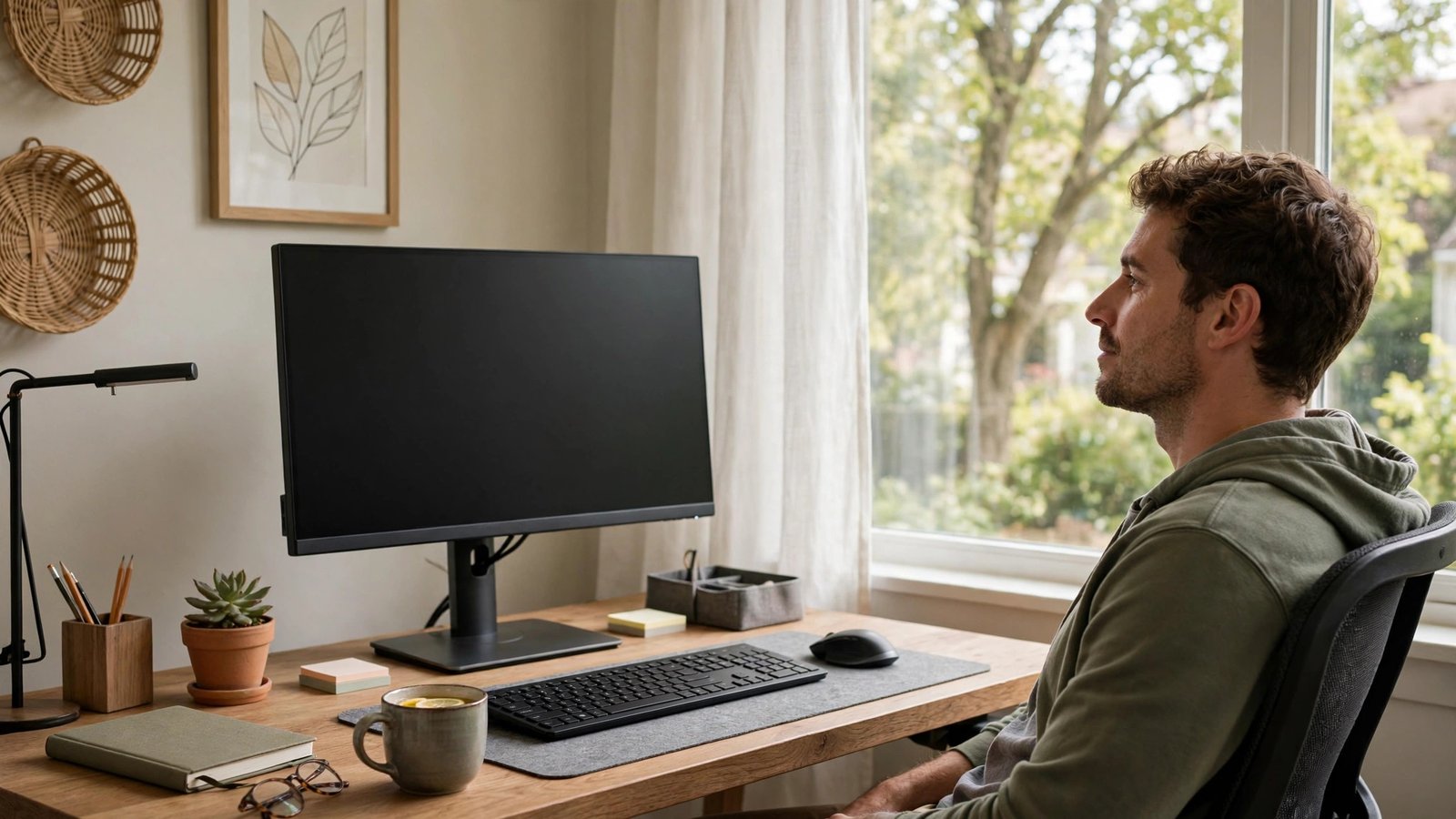
    Ten hours of screen time. By 4 p.m. your eyes burn, your vision blurs when you look across the room, and a headache is building behind your eyes. Digital eye strain — the cluster of symptoms from prolonged screen use — now affects the majority of office workers, and the standard workday keeps getting longer. The good news: eye strain is almost entirely a habits-and-setup problem, not an eyesight problem, and it responds to changes you can make today without buying anything. Here is the complete fix.

    Why Screens Wreck Your Eyes (It’s Not the Screen)

    The screen itself is not damaging your eyes — your behavior around it is. Three mechanisms dominate. First, blink rate collapse: normal blinking runs 15-20 times per minute; during concentrated screen work it drops to 5-7, and each missed blink lets the tear film evaporate, producing dryness, burning, and fluctuating blur. Second, sustained near focus: holding focus at 20 inches for hours fatigues the ciliary muscle inside the eye (the focusing muscle) exactly like holding a half-squat fatigues your thighs — the ache and distance-blur afterward are muscle fatigue, not damage. Third, setup sins: screens too close or too high force awkward gaze angles; glare and poor contrast make the eyes work harder for every character; and dry office air (often under 30% humidity) accelerates tear evaporation. None of this causes permanent eye damage — that is the reassuring part — but the daily misery is real and cumulative through the workweek. The American Optometric Association’s screen-time guidance details this mechanism officially.

    The 20-20-20 Rule and Its Upgrades

    The famous 20-20-20 rule — every 20 minutes, look at something 20 feet away for 20 seconds — works because it breaks all three strain mechanisms simultaneously: distance viewing relaxes the focusing muscle, the break restores normal blinking, and the pause interrupts the posture lock. But most people forget to do it, so make it automatic: set a recurring timer, use a break-reminder app (Stretchly, Big Stretch, and built-in OS reminders all work), or tie it to an existing habit (every time you finish an email, look out the window). Upgrade the rule for 10-hour days: every hour, take a full 5-minute break — stand, walk, look far away, blink deliberately. The deliberate blinking matters: 10 slow, complete blinks (fully closing the lids, not half-blinks) during each break re-coats the tear film more effectively than passive rest. Palming — covering closed eyes with warm palms for 30 seconds in a darkened break — relaxes both the eye muscles and the surrounding facial tension. These micro-breaks cost roughly 25 minutes across a 10-hour day and return far more in afternoon productivity than they consume.

    Fix Your Setup: Distance, Height, and Light

    Ergonomics do half the work permanently. Distance: the screen should be 20-28 inches from your eyes (about arm’s length) — closer forces excessive focusing effort; much farther forces squinting. Height: the top of the screen at or slightly below eye level, so your gaze angles slightly downward (10-20 degrees) — this position exposes less eye surface (reducing tear evaporation) and keeps neck neutral. Text size: if you are leaning in or squinting, the text is too small — increase zoom to 125-150% rather than punishing your eyes; there is no prize for tiny fonts. Glare: eliminate reflections on the screen — reposition relative to windows (perpendicular, not facing or backing them), close blinds as needed, and consider a matte screen filter for harshly lit offices. Ambient light should roughly match screen brightness — a glowing screen in a dark room forces constant pupil adjustment; a screen dimmer than bright surroundings washes out. Most monitors ship at retina-searing factory brightness — turn it down to match the room. Clean the screen weekly; dust and smudges scatter light and degrade contrast, making eyes work measurably harder. Our health section has more workstation wellness guides.

    Hydrate Your Eyes: Beating the Dryness

    For many 10-hour screen workers, dryness is the dominant symptom, and it has specific fixes. Artificial tears (preservative-free single-use vials for frequent use — preserved bottles irritate with overuse) used 2-4 times through the workday directly replace the evaporated tear film; keep them at your desk and use them on schedule, not just when burning starts. A warm compress for 5 minutes in the evening (warm washcloth over closed eyes) melts the oils in your eyelid glands, improving tear quality — particularly effective if your eyes feel gritty. Office air is the enemy: a small desktop humidifier, or even positioning away from HVAC vents blowing directly at your face, measurably reduces evaporation. Contact lens wearers suffer disproportionately — consider glasses for the longest screen days, since contacts divide the tear film and worsen dryness; if you must wear contacts, the artificial tears schedule becomes essential. Omega-3 supplements show modest evidence for dry eye improvement over weeks. And the simplest: conscious blinking — most screen workers can halve their dryness symptoms just by noticing their blink rate and correcting it during the 20-20-20 breaks.

    When Eye Strain Needs an Eye Doctor

    Habit and setup fixes resolve most digital eye strain — but not all, and some symptoms need professional evaluation. Book an eye exam if: strain persists despite two weeks of proper breaks, ergonomics, and dryness management; you have headaches centered behind the eyes most workdays; distance vision stays blurry for a long time after screen sessions; you are over 40 and have never had a presbyopia evaluation (the focusing fatigue of uncorrected presbyopia masquerades as screen strain); one eye is consistently worse; or you experience double vision, flashes, new floaters, or eye pain (distinct from strain ache — pain warrants prompt evaluation). The exam may reveal an outdated prescription, uncorrected astigmatism (which forces extra focusing effort at screens specifically), binocular vision issues, or dry eye disease needing prescription treatment. “Computer glasses” — single-vision lenses optimized for screen distance — help many workers, but get them prescribed after an exam, not from a drugstore guess. Blue-light glasses: the evidence for strain reduction is weak (the strain is behavioral, not spectral), though some people find the slight tint subjectively comfortable — spend the money on an exam instead. If screen symptoms worry you, reach out — and schedule the exam.

    Ten-hour screen eye strain comes from collapsed blink rates, sustained near-focus muscle fatigue, and poor setup — not screen damage. Break it with 20-20-20 plus hourly 5-minute breaks, position the screen at arm’s length slightly below eye level, kill glare, use preservative-free artificial tears, and humidify dry office air. Persistent strain despite two weeks of fixes deserves an eye exam.

    Conclusion

    Digital eye strain is a design problem with a design solution: break the focusing lock regularly, restore blinking, set up the workstation to work with your eyes instead of against them, and manage dryness directly. Implement the full protocol for two weeks and the 4 p.m. burn-and-blur should fade dramatically. Your eyes are not deteriorating from screens — they are asking for the breaks and setup they were designed to have. Give them that, and ten-hour days get comfortable again. Learn more about our practical health coverage. The National Eye Institute’s eye health resources cover digital eye strain prevention.

    Frequently Asked Questions

    Can screens permanently damage your eyes?

    No — extensive research finds no evidence that screen use causes permanent eye damage or worsens prescriptions. Digital eye strain is real and miserable but reversible; the symptoms resolve with breaks and proper setup. The concern is comfort and productivity, not lasting harm.

    Do blue-light glasses help with eye strain?

    Evidence is weak — most eye strain comes from blink reduction and focusing fatigue, not blue light wavelengths. Some users report subjective comfort from the tint. An eye exam and ergonomic fixes deliver far more proven benefit per dollar.

    How often should I use artificial tears?

    For screen-related dryness, 2-4 times through the workday on a schedule works better than waiting for burning. Use preservative-free single-use vials if applying more than 4 times daily — preservatives in multi-use bottles irritate with frequent use.

    Why do my eyes blur when I look up from the screen?

    That is focusing-muscle fatigue — the ciliary muscle locks into near-focus after hours and is slow to release for distance. It is the eye equivalent of muscle stiffness after holding a pose, and it resolves with distance-viewing breaks. Persistent prolonged blur deserves an exam.

    Are dark mode and night shift helpful?

    Modestly — they reduce overall light exposure and may improve comfort in dim environments, especially evening use. But they do not address the core mechanisms (blinking, focusing fatigue). Use them as comfort tweaks alongside the real fixes, not instead of them.

  • What Should I Do If I Miss a Dose of My Blood Pressure Medication?

    What Should I Do If I Miss a Dose of My Blood Pressure Medication?

    You were rushing out the door and forgot your blood pressure pill. Or you found yesterday’s pill still sitting in the organizer. Now you are wondering: take it now? Skip it? Double up tomorrow? This is one of the most common medication questions in America — and the answer is reassuringly simple for most blood pressure drugs, with a few important exceptions. Here is exactly what to do, what not to do, and how to stop missing doses in the first place.

    The General Rule: Take It When You Remember (With One Exception)

    For most blood pressure medications, the rule is straightforward: take the missed dose as soon as you remember, unless it is almost time for your next dose — in which case, skip the missed one and resume your normal schedule. “Almost time” generally means within a few hours of the next dose (for once-daily meds, if you remember the next morning, just take that day’s dose normally). Never double up — taking two doses at once can drop your blood pressure too far, causing dizziness, fainting, or in extreme cases dangerous hypotension. A single missed dose of most modern blood pressure medications (ACE inhibitors like lisinopril, ARBs like losartan, calcium channel blockers like amlodipine, thiazide diuretics like hydrochlorothiazide) is a minor event: these drugs work over days and weeks, not hours, and one missed dose barely moves your average pressure. The danger is not the single miss — it is the pattern. Missing doses a few times a month measurably worsens blood pressure control and raises cardiovascular risk; the occasional forgotten pill is noise, but habitual missing is signal. The American Heart Association’s medication guidance emphasizes this adherence-over-perfection principle.

    The Exceptions: Drugs You Must Not Miss

    Most blood pressure drugs are forgiving, but some are not. Beta-blockers (metoprolol, atenolol, propranolol) deserve special caution: stopping them abruptly — including via several missed doses — can cause rebound effects: racing heart, spiking blood pressure, chest pain, and in people with heart disease, genuinely dangerous cardiac events. If you miss beta-blocker doses, contact your doctor or pharmacist for guidance rather than just resuming; you may need a tapered restart. Clonidine is the other critical one: abrupt discontinuation causes severe rebound hypertension — agitation, headache, dangerously high pressure — and missed doses should prompt a call to your prescriber. Combination: if you take multiple blood pressure drugs and have missed several days of all of them, do not simply restart the full regimen at full dose — call your doctor, because restarting everything at once after a gap can overshoot. The principle: the longer-acting and more autonomically active the drug, the less forgiving the miss. Know which category your medications fall into — ask your pharmacist to flag any of yours as “do not miss” drugs. Our health guides cover more medication safety topics.

    What to Do Right Now: A Decision Tree

    Missed a dose? Walk through this. Step 1: Identify the drug — is it a beta-blocker, clonidine, or another “do not miss” medication? If yes, call your doctor or pharmacist now for instructions. Step 2: For standard blood pressure drugs — how long ago was the dose due? If less than half the dosing interval has passed (under ~12 hours for once-daily meds), take it now. If more than that, skip it and take the next dose on schedule. Step 3: Never take two doses together to “catch up.” Step 4: Note the miss — jot it in your phone or pill organizer log, because patterns matter more than incidents, and your doctor needs accurate adherence information (not the optimistic version). Step 5: Check how you feel — a single missed dose rarely causes symptoms, but if you feel dizzy, have chest pain, severe headache, or vision changes, seek care promptly rather than attributing everything to the missed pill. Step 6: If you have missed multiple consecutive days, call your prescriber before resuming — especially with beta-blockers or multiple medications. When in doubt, your pharmacist is the fastest expert available — a two-minute call resolves most missed-dose questions definitively.

    Stop Missing Doses: Systems That Actually Work

    Willpower is the worst adherence strategy; systems are the best. The evidence-backed approaches: link the pill to an unmissable daily anchor — morning coffee, brushing teeth, the evening news. The habit-stack (“after I pour coffee, I take my pill”) outperforms reminders alone. Use a weekly pill organizer — the visual “did I take it?” check eliminates the most common miss pattern (uncertainty leading to skipped doses). Set a phone alarm labeled with the drug name, and do not dismiss it until the pill is swallowed. Keep a backup dose where you will need it — desk drawer at work, travel bag — because “I left it at home” is a top miss reason. Use pharmacy auto-refill and 90-day supplies to eliminate the “ran out” gap, which causes more missed doses than forgetfulness. Ask your doctor about combination pills (two drugs in one tablet) if you take multiple medications — fewer pills means fewer misses. And be honest with your doctor about adherence — they cannot help with a problem they do not know about, and “I miss doses because of side effects” is fixable (different drug, different timing) while silent non-adherence just looks like treatment failure. If medication routines confuse you, reach out — though your pharmacist remains the best personal resource.

    For most blood pressure medications, a missed dose means: take it when you remember unless the next dose is near, then skip — never double up. Single misses barely matter; patterns do. Exceptions: beta-blockers and clonidine need prescriber guidance after misses (rebound risk), and multi-day gaps of any regimen warrant a call before restarting.

    Conclusion

    A missed blood pressure pill is a minor event for most medications — take it late or skip it, never double, and focus your energy on the system that prevents the next miss rather than guilt over this one. Know whether any of your drugs are in the unforgiving category (beta-blockers, clonidine), keep your pharmacist’s number handy, and be honest with your doctor about adherence. Blood pressure control is a long game won by consistency, not perfection. Learn more about our practical health coverage. The FDA’s medication adherence resources offer practical tools for staying on schedule.

    Frequently Asked Questions

    Can I take two pills tomorrow to make up for today?

    No — never double up on blood pressure medication. Doubling can drop pressure dangerously low, causing dizziness, fainting, or worse. Take the next scheduled dose normally and move on; one missed dose has minimal effect on overall control.

    What if I missed several days in a row?

    Call your doctor or pharmacist before resuming, especially with beta-blockers (rebound risk) or multiple medications. They may want you to restart gradually or be monitored. Do not silently restart a full regimen after a long gap.

    Will one missed dose raise my blood pressure dangerously?

    Extremely unlikely with standard medications — they work over days to weeks, and a single miss barely affects average pressure. The exception is abrupt beta-blocker or clonidine discontinuation, which can cause rebound effects. If you feel unwell, seek care regardless.

    Should I tell my doctor I miss doses?

    Yes — always. Doctors frequently intensify treatment (adding drugs, raising doses) for what looks like resistant hypertension but is actually non-adherence, exposing you to unnecessary medication. Honesty about misses leads to better solutions: simpler regimens, different timing, or combination pills.

    Does it matter what time of day I take blood pressure medication?

    Consistency matters more than the specific hour — same time daily keeps levels steady. Some evidence favors bedtime dosing for certain drugs, but the best time is the time you will reliably remember. Anchor it to an existing daily habit.